
It is no secret that Australia has the highest incidence of melanoma in the world. Incidence rates for melanoma were projected to more than triple between 2004 and 2021, thanks to improvements in detection tools and an increased awareness of skin cancer among other reasons.
Dermatologist Professor H. Peter Soyer relocated to Australia in the year 2007. The fact that Australia has such high rates of melanoma added to the importance of this professional move. Today, he is the Inaugural Chair of Dermatology at the University of Queensland.
London Agency had the opportunity to speak to Professor Soyer about the ground-breaking, world-leading research into melanoma that he has embarked upon.
The Australian Centre of Excellence in Melanoma Imaging and Diagnosis (ACEMID), a research platform of the University of Queensland, The University of Sydney and Monash University, received a major infrastructure grant of $10 million from the Australian Cancer Research Foundation in 2018. This infrastructure grant was followed by a National Health and Medical Research Council (NHMRC) grant awarded to Monash University in 2021 to screen a total of 15,000 study participants across 15 sites using innovative 3D total-body photography technology.
The objective of the study is to confirm that artificial intelligence (AI)-supported whole-body photography can improve melanoma diagnosis and avoid unnecessary diagnostic excisions (procedure involving cutting around and removing a skin lesion through the full thickness of skin). The study has already recruited over 4,800 people to participate at ACEMID 3D imaging sites in Queensland, New South Wales and Victoria, and the study protocol was recently published in the British Medical Journal.
To diagnose patients with melanomas, dermatologists usually conduct direct visual skin examinations combined with dermoscopy, a non-invasive imaging technique, and also rely on their clinical memory recall and the use of 2D digital images of skin lesions if available. However, the process to compose a patient body map via 2D imaging is time-consuming and not routinely done.
 |
According to Professor Soyer, melanoma overdiagnosis is a challenge. He said: “We have 17,000 invasive melanomas every year in Australia and 28,000 in situ (stage 0) melanomas; diagnostic drift in the diagnostic threshold over time could be responsible.” |
The ACEMID study participants attend visits according to their melanoma risk category (very high risk, high risk or low/average risk), every six, 12 and 24 months respectively, over three years. Participants undergo 3D total-body photography, dermoscopy imaging at study visits, and a baseline questionnaire is used to collect sociodemographic, phenotypic, quality of life and sun behaviour data. A follow-up questionnaire is administered every 12 months to capture any changes in sun behaviour and quality of life data. A saliva sample is also collected at the first visit for genetic testing.
Professor Soyer explained: “There are particular genes that we know signify a very high risk for melanoma and these can be detected from the saliva.”
The 3D total-body photography technology used in the study is manufactured by Canfield Scientific Inc (Parsippany, NJ, US), and captures 92 images which are then collated into a personalised avatar by an algorithm. Each 3D avatar comes in at a whopping 1.5GB of data, thanks to the high resolution used to get the clearest pictures.
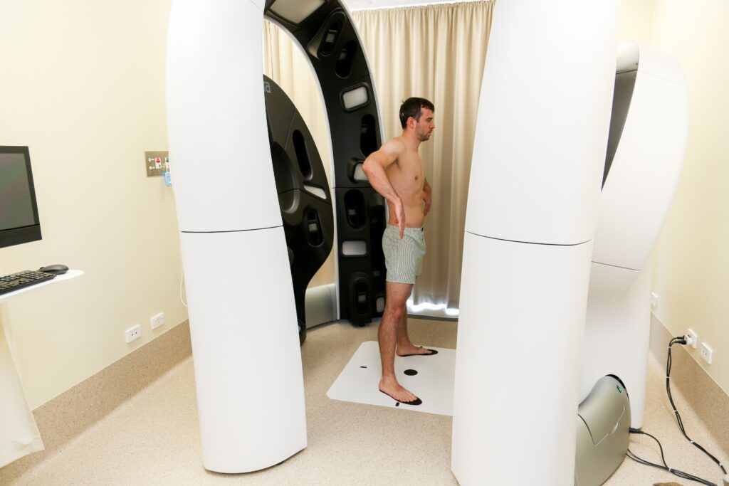
“It looks a little bit like a spaceship, but then there are 92 cameras. You go into it, you stand there, close your eyes, and then within a second, all the cameras are shooting and taking images.
“It’s not just the first study of its kind in Australia, it’s the first of its kind worldwide. Currently there are 77 of these systems installed worldwide: 23 in Australia, and 15 of these are part of our ACEMID research program.”
And so far, the results have been promising. Professor Soyer said: “We have diagnosed melanomas, which might have been otherwise missed.”
The study will not be completed for a few more years, as the participants need to be photographed several times over a three-year period and most study sites are still recruiting research participants.
Professor Soyer sees AI having an important role in the future of dermatology: “I’m not sure if this will be in five or 10 years, but there will be imaging centres with 3D or 2D machines fully integrated with artificial intelligence and run by skin imaging technicians. And dermatologists will be supported by AI in the decision-making process.
“I think the workflow that we have today will completely change and it will also open the door for telemedicine, not just for skin cancer, but also for skin rashes, such as psoriasis or eczema.
“I feel strongly that the virtual dermatologist will sooner than later become a reality.”
